Swabian Joint Center
The term endoprosthetics refers to the replacement of a severely diseased joint with an artificial joint (an endoprosthesis).
Prof. Dr. med. Michael Maier and his endoprosthetics team at SGZ are specialists in the field of shoulder, hip and knee endoprosthetics.
Joint replacement of hip and knee is one of the most common operations in Germany. The estimated number of hip and knee joints implanted in Germany is around 450,000 per year, with approximately 250,000 hip and 200,000 knee prostheses being fitted. These figures have remained stable in recent years.
The annual number of artificial joint implantations in Germany is significantly lower with about 25,000 endoprostheses per year, and the trend is increasing. Prof. Maier is an expert in the installation of anatomical shoulder prostheses in patients with shoulder arthrosis and inverse shoulder prostheses in patients with defect arthropathy and complex, non-reconstructable fractures of the humeral head.
Endoprosthesis operations are very successful operations; hip joint replacement has even been ennobled by the renowned medical journal "The Lancet" with the title "operation of the century".
Professor Maier, is the typical endoprosthetics patient more driven by his or her level of suffering, or are there now also preventive interventions for only minor problems but foreseeable complaints?
The typical patient who comes to the Swabian Joint Center for joint replacement surgery has experienced a significant reduction in quality of life in recent years due to arthrosis (wear and tear of cartilage), and the level of suffering is correspondingly high.
While at the beginning of the disease, conservative therapy with drug therapy, weight normalization, physiotherapy and exercise therapy, hyaluronic acid and autologous blood therapy provided relief, these forms of therapy often no longer help the patient with advanced arthrosis.
Preventive surgical interventions only make sense if the patient has incipient arthrosis with a known cause, such as axial defects in the knee with incipient bowleg arthrosis or incipient femoral head necrosis. Preventive interventions can be useful here.
If the progression of the arthrosis disease has led to complete wear and tear of the joint cartilage, conservative therapy often does not provide any further relief. This patient is driven by a pronounced level of suffering and comes to the doctor's office with severe joint pain. He often had to limit his individual activities of daily life, which can affect his beloved sport of golf, for example.
Here, the positive effect of sport on health must be weighed against the surgical risk of a joint replacement and its chances of success. If you look at the preventive effect of physical activity, it has been proven that 150 minutes of moderately intensive sporting activity significantly reduces the risk of cardiovascular disease, stroke and type 2 diabetes. The requirement for a joint replacement of the shoulder, hip or knee joint must therefore be to enable the athletically active patient who has remained young and wants to keep fit through sports to be able to practice sports again without pain through the artificial joint.
Professor Maier, when does the physician have a basis for deciding whether to replace the joint partially or completely?
The indication for a joint replacement of the shoulder, hip or knee must always be based on a comprehensive medical history, a clinical examination and appropriate radiological diagnostics. The correct indication is decisive for the success of the therapy for each individual patient.
During the clinical examination, the joint mobility of the affected joint is assessed in a side comparison, structures close to the joint such as muscles and tendons are tested for functionality and special pain and stability tests are applied. The radiological diagnostics must take into account the particularity of the joint in each case and includes X-rays, special X-ray stress images supplemented by magnetic resonance imaging and, if necessary, computer tomography. On the basis of these imaging diagnostics, the joint specialist sees the typical signs of osteoarthritis such as joint space narrowing, joint deformities and marginal attachments. Only when these objectifiable criteria are available and match the patient's personal level of suffering is the basis for the decision to replace the affected joint completely or partially.
Severe arthrosis using the example of the hip joint
Prof. Maier, what happens for patients after endoprosthetic operations? Do you have any approximate timescales that could be used for a good healing process?
After the joint replacement, rehabilitation begins on the day of the operation with the aim of integrating the artificial joint into everyday life as quickly as possible. With hip and knee prostheses, patients are immediately allowed to put full weight on the joint and training is carried out under physiotherapeutic supervision with the aim of achieving good muscular control of the joint with normalisation of the gait pattern.
For the overhead athlete with shoulder prosthesis, the cementless prosthesis must be integrated into the bone in a stable manner so that the load does not cause loosening. This should be possible three months after the operation if the healing process is good.
In general, low-impact sports are recommended throughout endoprosthetic operations.
In the case of an anatomical shoulder joint replacement, patients can begin with overhead sports such as golf again after 6 months if the healing process is good. Surprisingly, studies among experienced golfers with shoulder prostheses have shown no problems at all with resuming their sport. The good news for all golfers is that an optimally functioning prosthesis can even improve the handicap as well as slightly improve the length of the stroke!".
Artificial shoulder joint
Professor Maier, the shoulder is a special joint with a large range of motion and is very demanding, especially for overhead athletes. What is so special about shoulder endoprosthetics?
The shoulder is a very important joint, especially for the overhead athlete. The large radius of movement that our hand needs for optimal use is ensured by the design of the arm as a link chain and the special mobility of the shoulder joint and shoulder girdle.
The special feature of the ball joint shoulder is that the glenoid cavity is small compared to the ball, the humeral head. Think of it like comparing golf tee to golf ball. Because the glenoid cavity is small, stabilisation at the shoulder joint requires the strength of a well-trained musculature, the so-called rotator cuff, which is why the shoulder is also referred to as a "friction-locked" joint.
The rotator cuff cannot be palpated because it is located deep below the shoulder cap muscle (deltoid muscle). The muscles of the rotator cuff move from the shoulder blade to the head of the humerus, where they attach their tendons and stabilise the head of the humerus in the small socket at the shoulder joint, which is an essential component of pain-free mobility of the upper arm in the shoulder joint. This applies to the natural shoulder as well as to the artificial joint.
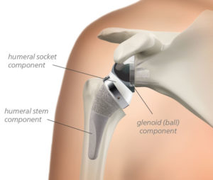
In recent years, artificial joint replacement of the shoulder has become increasingly important. Good long-term results of established implant systems have strengthened the confidence in the known shoulder prostheses, which is confirmed by the constantly increasing number of operations. A decisive factor for the success of a shoulder endoprosthesis operation is that the surgeon selects the right prosthesis model for the respective shoulder disease. If an anatomical total shoulder endoprosthesis is fitted in the case of shoulder arthrosis, the rotator cuff mentioned above must be functionally intact, otherwise premature loosening will occur.
Anatomical, shaft-free shoulder prosthesis
In young patients with necrosis of the humeral head and a regular socket, only a replacement of the diseased humeral head can be made, a so-called hemiprosthesis.
The situation is different in the clinical picture of defective arthropathy, where a destroyed, non-reconstructable rotator cuff is the main problem. In this case, a normal anatomical shoulder prosthesis must not be fitted, as this would lead to premature loosening. Fortunately, today we have the inverse shoulder prosthesis as a successful therapy method.
Inverse shoulder prosthesis
Current movement analysis studies have shown that endoprosthetic replacement of the shoulder leads to a significant improvement in the maximum range of movement when performing everyday movements, which, along with pain reduction, is the decisive parameter for patient satisfaction.
- Shoulder arthrosis (omarthrosis) with final wear and tear of the joint cartilage, when conservative therapy no longer brings you any relief.
- Destruction of the shoulder joint through rheumatoid arthritis
- Necrosis of the humeral head (death of the humeral head) e.g. after prolonged cortisone treatment
- Complicated fracture of the humeral head without the possibility of reconstruction (fracture prosthesis)
- Unsuccessful fracture treatment of the head of humerus with necrosis of the head of humerus
- Disease pattern of defect arthropathy, which means that a pronounced arthrosis of the shoulder has developed after a massive irreparable defect of the rotator cuff. If you as a patient with this clinical picture have massive pain and a pronounced loss of shoulder function, the so-called inverse shoulder prosthesis is an excellent treatment option.
- At the shoulder joint, both the humeral head and the socket can be replaced artificially.
- In most cases, the humeral head and the socket are replaced.
- This is known as a total shoulder endoprosthesis (shoulder TEP).
- Yes, if the socket is not affected, for example in the case of necrosis of the humeral head, only a replacement of the diseased humeral head, a so-called hemiprosthesis, can be made.
- In a defective arthropathy, shoulder arthrosis occurs due to a destroyed, non-reconstructable rotator cuff with subsequent joint destruction. In this case, no normal anatomical shoulder TEP may be inserted, as this would lead to premature loosening.
- Nowadays, the inverse shoulder prosthesis is used in this clinical picture.
- The inverse prosthesis works excellently because it creates a fixed rotation centre with congruent joint surfaces, which improves stability in the case of defect arthropathy.
- Shoulder joint replacement is performed in Germany about 10 times less frequently than hip joint replacement.
- For this reason, it is essential that the presentation be made at a specialised shoulder centre before the operation.
- It has been scientifically proven that the surgeon who performs the procedure frequently and routinely with his team has better results and a lower complication rate.
- Patients are hospitalized for about 4 days after shoulder joint replacement. During the inpatient stay, targeted physiotherapy begins.
- The loss of pain and the improvement of shoulder function are usually so good after shoulder joint replacement that sporting activity can also be resumed after intensive rehabilitation.
- Fitness, Nordic walking, hiking, tennis and golf are among the possible sports.
- Full mobility of the shoulder is usually restored after 3 months.
- We recommend clinical and radiological follow-up according to 1, 3, 5 years and then every 5 years.
Artificial hip joint
Professor Maier, which surgical techniques are currently the best for the hip? One always reads about minimally invasive or AMIS. Can you explain the differences in a few words?
Minimally invasive approaches to the hip joint have become more and more popular in recent years due to the increasing demand of patients to have the shortest possible hospital stay and quick rehabilitation. While 20 years ago, you as a patient still had inpatient stays of 2 weeks after a hip joint replacement with a classic approach, this has recently been reduced to a few days.
In order to enable short hospital stays, classical surgical techniques, which often required muscle detachment, were abandoned and replaced by minimally invasive approaches. These approaches respect the course of the musculature and tendon attachments and aim to minimise damage to muscles close to the joint. The AMIS approach is the anterior minimally invasive approach in which a natural muscle gap to the hip joint is used in a very elegant way. Muscles, tendons, vessels and nerves are pushed to the side.
So it is not the small skin incision that is crucial in AMIS access because it is not the skin that can make life difficult for patients after hip replacement surgery. It is the severed or injured muscles and tendons of the large muscles surrounding the hip joint that, once they are severed or injured, heal slowly and severely, sometimes not at all. The clear advantage of minimally invasive approaches is therefore that muscles, tendons and mechanoreceptors are preserved. The mechanoreceptors are important for our depth sensitivity and thus for a stable gait pattern. If we leave these structures intact during prosthesis implantation, the patient can start rehabilitation earlier after the procedure, the hospital stay is shortened and the patient returns more quickly to his or her individual daily activities such as golf.
Professor Maier, what about the materials there? What experience has been gained in the meantime? There is nothing worse than to read at the breakfast table after 10 years that the type of metal from 10 years ago is often defective...
In addition to the correct surgical method, the material and thus the durability of the respective prosthesis is decisive for a good long-term result. Here it is important to remember that modern hip prostheses are assembled from individual parts according to the modular principle: Acetabular cup, cup insert (inlay), ball head and hip stem.
Components of a hip endoprosthesis
During the operation, the modified femoral head including part of the neck of the femur is removed and then, after the modified acetabulum has been milled, an artificial cup is inserted into the pelvis and a prosthesis stem into the femur. Modern prostheses are made of different metal alloys (titanium, cobalt-chrome).
If the bone quality is good, cementless short shaft prostheses made of titanium alloys are often used nowadays. These are easy to implant minimally invasively and initially jam in the bone, which we call primary stability. Since the titanium surface is porous, the bone grows within a few months and fixes the prosthesis additionally, which is called secondary stability. In older patients with softer bone, e.g. due to osteoporosis, the cemented prosthesis is a very good solution.
The inlay is inserted into the socket as a running surface and the prosthesis head is attached to the prosthesis shaft. The movement then takes place between these two prosthesis parts, which we call sliding pairing. In the past, metal heads and polyethylene inlays were often used as sliding pairings. Due to the high abrasion of the polyethylene inlays at that time, however, strong abrasion-induced foreign body reactions occurred in many cases, which led to local bone dissolution with subsequent prosthesis loosening and resulted in alternating operations. The same problem occurred with hip cap prostheses, where a metal-metal sliding pairing was used. Metal-metal sliding pairings should no longer be used today due to the massive metal abrasion.
Instead, we now prefer modern ceramic-polyethylene and ceramic-ceramic sliding combinations, which have to meet ever higher demands due to the increased level of activity of our young patients. Therefore, the material properties of these pairings have been further improved in order to have the lowest possible abrasion of the pairing surfaces, which led to the development of modern ceramics and modern ultra-high cross-linked polyethylene with vitamin E. Today we have ideal conditions and a durability of more than 25 years is normal nowadays.
- In the case of hip arthrosis (coxarthrosis) when there has been a final wear and tear in the hip joint and conservative therapy no longer provides relief.
- Dysplasia coxarthrosis
- femoral head necrosis
- Femoral neck fracture
- At the hip joint, the femoral head and the acetabulum can be replaced artificially.
- If both components are replaced, this is called a total hip endoprosthesis (hip TEP).
- An individual operation planning is carried out for each patient before the operation.
- Most of the hip joint replacement operations can be carried out at the Swabian Joint Centre using minimally invasive surgical techniques that are gentle on the muscles. This shortens the post-treatment time, patients become mobile faster and the final results are also better due to the avoidance of muscle damage.
- The implantation of an artificial hip joint is one of the most successful operations with a patient satisfaction rate of over 90%.
- The hip prosthesis team around Professor Maier has specialised in the minimally invasive AMIS approach for the implantation of a hip TEP. Compared to conventional hip surgery techniques, the AMIS technique is less traumatic because the muscles around the hip are not severed but only pushed to the side.
- We will advise you comprehensively and individually on the extent to which the AMIS technique for implantation of an artificial hip can be carried out on you, so that your quality of life improves again quickly.
- Due to the strong muscle mantle of the hip, the artificial hip joint replacement is a demanding hip operation, which should be performed by experienced, certified hip surgeons if possible.
- It has been scientifically proven that the surgeon who performs the procedure frequently and routinely with his team has better results and a lower complication rate.
- Patients are hospitalized for about 4 days after hip joint replacement. During the in-patient stay, targeted physiotherapy begins.
- Fitness, Nordic walking, hiking, swimming and cycling are among the recommended sports.
We recommend clinical and radiological follow-up after 1, 3, 5 years and every 5 years thereafter
- In the case of hip arthrosis (coxarthrosis) when there has been a final wear and tear in the hip joint and conservative therapy no longer provides relief.
- Dysplasia coxarthrosis
- femoral head necrosis
- Femoral neck fracture
- At the hip joint, the femoral head and the acetabulum can be replaced artificially.
- If both components are replaced, this is called a total hip endoprosthesis (hip TEP).
- An individual operation planning is carried out for each patient before the operation.
- Most of the hip joint replacement operations can be carried out at the Swabian Joint Centre using minimally invasive surgical techniques that are gentle on the muscles. This shortens the post-treatment time, patients become mobile faster and the final results are also better due to the avoidance of muscle damage.
- The implantation of an artificial hip joint is one of the most successful operations with a patient satisfaction rate of over 90%.
- The hip prosthesis team around Professor Maier has specialised in the minimally invasive AMIS approach for the implantation of a hip TEP. Compared to conventional hip surgery techniques, the AMIS technique is less traumatic because the muscles around the hip are not severed but only pushed to the side.
- We will advise you comprehensively and individually on the extent to which the AMIS technique for implantation of an artificial hip can be carried out on you, so that your quality of life improves again quickly.
- Due to the strong muscle mantle of the hip, the artificial hip joint replacement is a demanding hip operation, which should be performed by experienced, certified hip surgeons if possible.
- It has been scientifically proven that the surgeon who performs the procedure frequently and routinely with his team has better results and a lower complication rate.
- Patients are hospitalized for about 4 days after hip joint replacement. During the in-patient stay, targeted physiotherapy begins.
- Fitness, Nordic walking, hiking, swimming and cycling are among the recommended sports.
- We recommend clinical and radiological follow-up after 1, 3, 5 years and every 5 years thereafter
Artificial knee joint
Prof. Maier, how is the progress in knee endoprosthetics in terms of surgical techniques in recent years to be assessed?
Nowadays, patients can also count on excellent durability for knee prostheses. Eight out of ten total knee endoprostheses today last for 25 years. If we compare the satisfaction of hip and knee prosthesis patients, 90% of patients are satisfied with the results after a hip joint replacement, compared to only 80% for the knee. In order to improve this value, a lot of research has been and is being done on the optimal implantation technique.
Total knee endoprosthesis
The challenge in knee joint replacement is that it is not a ball and socket joint like the hip, but rather the largest joint in the human body consisting of three partial joints, and these partial joints form a functional unit. The three partial joints must be taken into account when choosing the right joint replacement. Only then can an optimal result be achieved and the patient will ideally get a "forgotten knee" in addition to pain reduction and improved function, which means that he can use his new artificial knee joint in everyday life like the natural knee joint and forget that it is an artificial joint. The cause analysis of dissatisfied patients has shown that surgical factors such as implant alignment play a central role. If the artificial knee joint is installed with a rotation error, it cannot function, the patient suffers pain and has poor function.
This is where we discuss today which surgical technique is the best to install the artificial joint as precisely as possible. Until now, neither navigation nor patient-specific instruments have been able to show a significant superiority over conventional surgical techniques. Perhaps newer approaches such as kinematic alignment, which involves positioning the prosthesis strictly along the axes of rotation of the natural knee, will bring further improvement.
However, there is one important factor that we must not forget despite all the discussion about the progress of op techniques: The experienced surgeon who knows his trade and who reconstructs the knee joint kinematics optimally in three dimensions when fitting the knee joint is the decisive factor for success. Manual errors during prosthesis installation must be avoided at all costs - an experienced surgeon is more valuable - every new, seemingly groundbreaking surgical technique.
Prof. Maier, what about the design and materials of knee prostheses? Perhaps you can explain the term "partial preservation" to us in more detail? How does it work exactly? And what is a partial endoprosthesis?
The design and materials of knee prostheses have been continuously improved in recent years, so that today, individual adaptation to the patient with maximum preservation of the non-damaged parts of the joint has become possible. The materials are now so good that they can last well over 20 years if correctly fitted.
In the past, the material was often suspected when patients had problems with their knee prosthesis. Today we know that it is not so much the material that fails but rather the surgeon. Studies show that manual errors such as rotation errors lead to poor results, especially in the knee joint.
The successful operation begins outside the operating room with an exact op planning including axis alignment, angle measurement, determination of the type of prosthesis and determination of the prosthesis size.
When selecting a prosthesis, the aim is to preserve as much of the body's own tissue structure as possible using the smallest possible implant (partial preservation). Corresponding to the three partial joints of the knee joint, each part can nowadays be replaced separately, which makes sense if the disease is limited to this part of the joint only. This means that there is a partial endoprosthesis on the inside or outside (sled prosthesis) and the joint surface behind the kneecap can also be replaced in isolation.
The most common partial joint replacement is the inner sled prosthesis for bowleg arthrosis of the knee. In this case, the inner side of the knee joint is replaced with a prosthesis and the remaining joint including cruciate ligaments is retained. With the correct indication and a clean surgical technique, excellent long-term results and a high level of patient satisfaction can be expected. Compared to full prostheses on the knee, partial prostheses are characterized by higher patient satisfaction, better mobility, lower surgical risks and a higher rate of "forgotten knees".
Inner sled prosthesis
- In the case of knee arthrosis (gonarthrosis) when there has been a final wear and tear in the knee joint and conservative therapy no longer provides relief.
- rheumatoid arthritis
- Symptomatic knee instability
- Posttraumatic Arthrosis
- Partial joint replacement means that, corresponding to the 3 partial joints at the knee joint, each part is replaced separately, which means that there is a half-sided prosthesis on the inside or outside (unicondylar joint replacement = sled prosthesis) and the joint surface behind the kneecap can also be replaced in isolation (isolated retropatellar replacement).
- The exact clarification prior to the operation is decisive here, as otherwise the results of the partial joint replacement may be poor.
- With the right indication and a clean surgical technique, excellent long-term results and high patient satisfaction can be expected.
- In cases of pronounced wear and tear of several compartments of the knee joint, a complete joint replacement (bicondylar joint replacement with patellar back surface replacement) must often be carried out.
- The medial Oxford sled prosthesis used at the Swabian Joint Center has very good long-term results documented in the literature with survival rates of 91% after 20 years.
- In addition to an excellent surgical technique, it is crucial that the indication is set correctly.
- The classic indication here is anteromedial gonarthrosis with complete cartilage loss.
- Classic indication for partial joint replacement (sled prosthesis) on the inside of the knee joint in bowleg arthrosis (varus gonarthrosis)
X-ray control after implantation of a sled prosthesis on the inside of the knee joint after bowleg arthrosis (varus gonarthrosis)
- Follow-up treatment begins on the day of the operation and is intensified during the approximately 4-day inpatient stay.
- Early mobilisation helps to strengthen the muscles surrounding the knee joint.
- With correct implantation and good muscular guidance, artificial joints last for more than 20 years.