Läsionen am Bizepsanker der Schulter sind eine häufige Ursache von Schulterschmerzen, insbesondere beim sportlichen Patienten der Überkopfsportarten wie Tennis, Golfen, Volleyball, Handball, Basketball ausübt: Prof. Dr. med. M. Maier vom Schwäbischen Gelenkzentrum und die Klinik für diagnostische und interventionelle Radiologie der Universitätsklinik Heidelberg erforschen, inwiefern diese Verletzungen mittels spezieller MRT-Diagnostik besser diagnostiziert werden können.
Diagnostic performance of 3D-multi-Echo-data-image-combination (MEDIC) for evaluating SLAP lesions of the shoulder
Background: Superior labral anterior to posterior (SLAP) lesions remain a clinical and diagnostic challenge in routine (non-arthrographic) MR examinations of the shoulder. This study prospectively evaluated the ability of 3DMulti-Echo-Data-Image-Combination (MEDIC) compared to that of routine high resolution 2D-proton-density weighted fat-saturated (PD fs) sequence using 3 T-MRI to detect SLAP lesions using arthroscopy as gold standard.
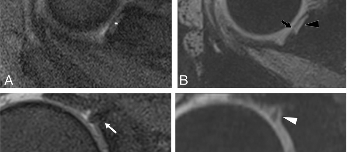
Methods: Seventeen consecutive patients (mean age, 51.6 ± 14.8 years, 11 males) with shoulder pain underwent 3 T MRI including 3D-MEDIC and 2D-PD fs followed by arthroscopy. The presence or absence of SLAP lesions was evaluated using both sequences by two independent raters with 4 and 14 years of experience in musculoskeletal MRI, respectively. During arthroscopy, SLAP lesions were classified according to Snyder’s criteria by two certified orthopedic shoulder surgeons. Sensitivity, specificity, positive predictive value (PPV) and negative predictive value (NPV) of 3D-MEDIC and 2D-PD fs for detection of SLAP lesions were calculated with reference to arthroscopy as a gold standard. Interreader agreement and sequence correlation were analyzed using Cohen’s kappa coefficient. Figure 1 demonstrates the excellent visibility of a proven SLAP lesion using the 3D-MEDIC and Fig. 2 demonstrates a false-positive case.
Results: Arthroscopy revealed SLAP lesions in 11/17 patients. Using 3D-MEDIC, SLAP lesions were diagnosed in 14/17 patients by reader 1 and in 13/17 patients by reader 2. Using 2D-PD fs, SLAP lesions were diagnosed in 11/17 patients by reader 1 and 12/17 patients for reader 2. Sensitivity, specificity, PPV, and NPV of 3D-MEDIC were 100.0, 50.0, 78.6, and 100.0% for reader 1; and 100.0, 66.7, 84.6, and 100% for reader 2, respectively. Sensitivity, specificity, PPV, and NPV of 2D-PD fs were 90.9, 83.3, 90.9, and 83.3% for reader 1 and 100.0, 83.3, 91.7, and 100.0% for reader 2. The combination of 2D-PD fs and 3D-MEDIC increased specificity from 50.0 to 83.3% for reader 1 and from 66.7 to 100.0% for reader 2. Interreader agreement was almost perfect with a Cohen’s kappa of 0.82 for 3D-MEDIC and 0.87 for PD fs.
Conclusions: With its high sensitivity and NPV, 3D-MEDIC is a valuable tool for the evaluation of SLAP lesions. As the combination with routine 2D-PD fs further increases specificity, we recommend incorporation of 3D-MEDIC as an additional sequence in conventional shoulder protocols in patients with non-specific shoulder pain.

